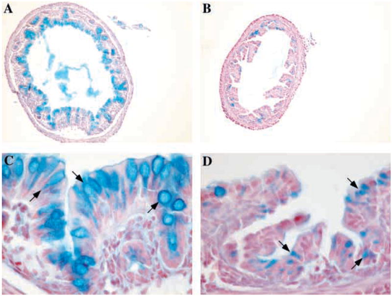
Fig. 4.

Klf4−/− mice showed a dramatic decrease in the number of goblet cells in the colon. (A–D) Alcian Blue staining for acidic mucins in the colon of postnatal day 1 mice. At low power, wild-type mice (A) had numerous Alcian Blue-positive cells, but very few Alcian Blue positive cells were seen in the Klf4−/−mice (B). At higher power, Alcian Blue staining was seen only in the goblet cells (arrows) of wild-type mice (C), while Klf4−/− mice (D) displayed numerous epithelial cells with small amounts of Alcian Blue staining material (arrows) but no normal goblet cell morphology.
