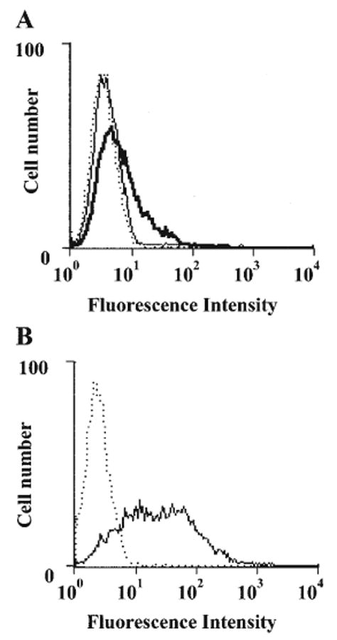
Figure 1.

FACS analysis of surface expression of CD26 and ecto-ADA on human gingival fibroblasts. (A) Human gingival fibroblasts (1 × 10⁶ cells) were incubated with (bold line) or without (solid line) Jurkat cell lysate for 30 min at 37°C, washed twice, and stained with anti-ADA plus FITC-donkey anti-goat IgG. Normal goat IgG was utilized as a control (dotted line). (B) Untreated human gingival fibroblasts were stained with PE-anti-human CD26 mAb (solid line) or isotype-matched murine myeloma protein (dotted line). The data shown are representative of 3 separate experiments.
