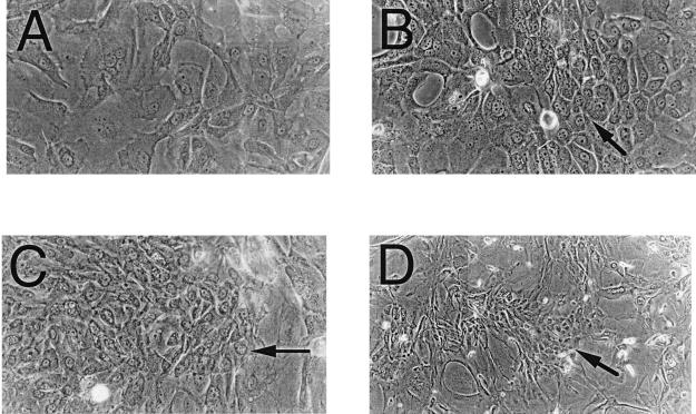
Figure 2.
Photomicrographs of Id-transfected HFKs under differentiating stimuli. Phase-contrast photomicrographs demonstrate focal regions of smaller, phenotypically less differentiated cells (arrows) in Id-1- (B), Id-2- (C), and Id-3- (D) transfected HFKs vs. vector control-transfected cells (A), which show a typical differentiated phenotype of large, flattened cells with increased cytoplasmic-to-nuclear ratios.

