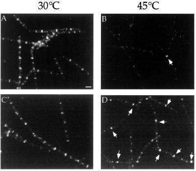
Figure 1.
Localization of FtsZ-GFP. Cells containing ftsZ-gfp were grown in LB to exponential phase (OD600 ≈ 0.5) at 30°C, then diluted 1:5 into fresh medium at 30°C (A and C) or 45°C (B and D). Samples were taken 45 min after dilution and analyzed by immunofluorescence microscopy by using primary antibodies against FtsZ and a Cy-3-conjugated secondary antibody. FtsZ rings appear as bands across the short axis of the cell. (A and B) PL642 (ftsZ-gfp) at 30°C (A) and after 45 min of growth at 45°C (B). Note that only a single ring of FtsZ is visible in this field of cells (arrow). (C and D) PL710 (ftsZ-gfp ezrA1) at 30°C (C) and after 45 min of growth at 45°C (D). Arrows indicate representative FtsZ rings. Bars = 1 μm.

