Figure 2.
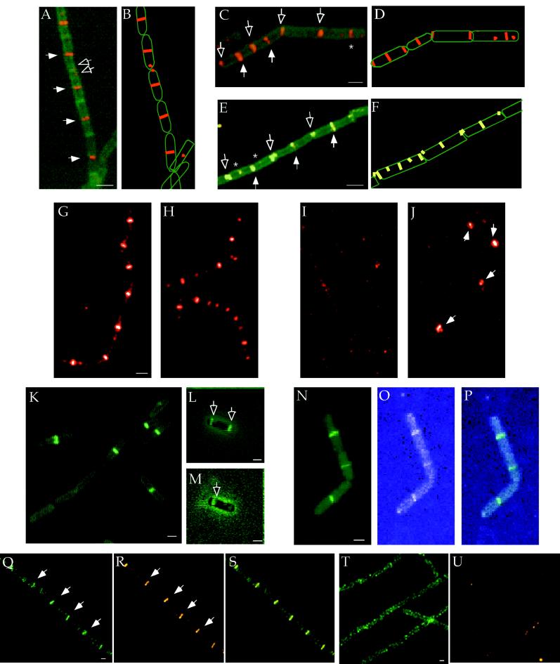
Localization of FtsZ and EzrA. (A–F) Localization of FtsZ in wild-type and ezrA mutant cells during exponential growth in LB at 37°C. (A, C, and E). FtsZ is stained red or yellow (because of the use of different filter sets) and the cell wall is stained green (see Materials and Methods.) Closed arrows indicate medial FtsZ rings. Open arrows point to polar rings or foci of FtsZ. Asterisks denote rings or foci of FtsZ at quarter positions. Merged images of FtsZ and cell walls (A, C, and E) and cartoons of the micrographs (B, D, and F). (A and B) Wild-type cells. Note the even spacing of the medially positioned FtsZ rings. Small foci of FtsZ are occasionally visible at the cell poles (open arrows). (C and D) ezrA1 ftsZ+ cells (PL730). The irregular spacing of the FtsZ rings in the ezrA− cells is because of FtsZ localization at polar and medial positions. Some cells have multiple FtsZ rings. The cell on the upper right has a single FtsZ ring at a quarter position. (E and F) ezrA∷spc, ftsZ+ cells (PL867). (G–J) Localization of FtsZ in ezrA+ (G and I) and ezrA mutant cells (H and J) before (G and H) and 100 min after (I and J) depletion of FtsZ. Strains PL919 (ezrA+ Pspac−ftsZ) and PL928 (ezrA∷spc Pspac−ftsZ) were grown in LB at 37°C in the presence of IPTG (1 mM to express ftsZ) and chloramphenicol (2 μg/ml to maintain the Pspac-ftsZ plasmid). At mid- to late exponential phase, cells were washed and diluted 1:10 in LB with chloramphenicol with (G and H) and without IPTG (I and J). FtsZ is immunostained red. The exposure times in G and H and in I and J are identical. Arrows point to rings of FtsZ in the ezrA mutant cells after FtsZ depletion. (K–M) Localization of EzrA-GFP in live cells, strain PL847, grown at 30°C. (K) EzrA-GFP in vegetatively growing cells sampled from solid rich medium. EzrA-GFP is distributed throughout the membrane and appears in some cells as a band or ring at midcell. (L and M) Localization of EzrA-GFP in cells 1.5 (L) and 3 (M) hr after the initiation of sporulation. Fluorescence and phase contrast images are superimposed. Open arrows point to polar rings of EzrA. (N–P) Colocalization of FtsZ-GFP and EzrA-BFP in live cells. Strain PL995 (ezrA-bfp ftsZ+ Pspac-ftsZ-gfp) was grown in LB at 30°C (5 μM IPTG) and sampled during exponential growth. (N) FtsZ-GFP. (O) EzrA-BFP. (P) Overlay of FtsZ-GFP (N) and EzrA-BFP (O). (Q–U) Localization of EzrA-GFP in the presence and absence of FtsZ. FtsZ depletion was carried out essentially as described above. Strain PL851 (ezrA-gfp Pspac-ftsZ) was grown in LB at 30°C in the presence (Q–S) or absence of IPTG (T and U). Samples were taken after approximately four mass doublings, fixed, and prepared for immunofluorescence microscopy. EzrA-GFP is immunostained green and FtsZ is immunostained red. (Q) EzrA-GFP. (R) FtsZ. (S) overlay of EzrA-GFP (Q) and FtsZ (R). (T) EzrA-GFP without IPTG, after depletion of FtsZ. Rings are no longer visible in these cells. Instead, only a punctate pattern of bright fluorescence is observed. (U) Residual FtsZ staining after depletion. The exposure time is the same as that in T. Very little FtsZ protein is present and ring formation is abolished. Bars = 1 μm.

