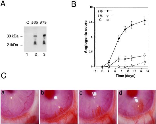
Figure 1.
Implanted Figf/Vegf-D-expressing cells induce neovascularization in rabbit corneas. (A) Figf/Vegf-D expressed in CHO cells. Equal volumes of culture supernatants from clones 65 and 79 were precipitated and analyzed by Western blot using an anti-Figf/Vegf-D rabbit polyclonal antiserum. (B) CHO cells (4 × 10⁴) expressing Figf/Vegf-D were surgically implanted into the corneas. New blood vessel growth was recorded every other day with a slit lamp stereomicroscope. Angiogenic scores were calculated on the basis of the number of vessels and their growth rate and plotted versus time (for experimental details see Materials and Methods). Angiogenic score data are the mean values obtained from the response scored in all animals in this study. C, CHO mock transfectant clone; #65, clone expressing low levels of Figf/Vegf-D (0.1 ng/ml protein in supernatant); #79 clone expressing higher levels of Figf/Vegf-D (approximately 0.5 ng/ml protein in supernatant). (C) Pictures of rabbit corneas from a representative experiment. (a) Corneal implant of CHO mock transfectant. Clone 79 promotes and sustains vascular growth over time at day 6 (b), 9 (c), and 14 (d). Corneas were photographed with a stereomicroscope. Magnification: ×18.

