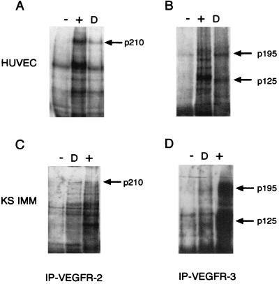
Figure 4.
Figf/Vegf-D-induced tyrosine phosphorylation of VEGFR-2 and VEGFR-3. HUVECs and KS-IMM cells were incubated with Figf/Vegf-D. After stimulation receptors were immunoprecipitated with antireceptor antibodies and analyzed by Western blotting with an antiphosphotyrosine mAb. (A and B) Phosphorylation of VEGFR-2 and VEGFR-3 in HUVECs. (C and D) Phosphorylation of VEGFR-2 and VEGFR-3 in KS-IMM cells. Positive control (+) and Figf/Vegf-D stimulation (D) is indicated. Arrows denote the position of the phosphorylated 210-kDa VEGFR-2 protein and the positions of the phosphorylated, proteolytically processed 125-kDa and unprocessed 195-kDa forms of VEGFR-3.

