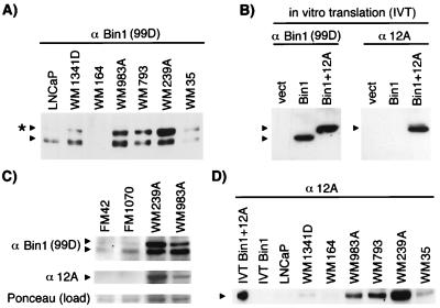
Figure 3.
Western blot analysis of Bin1 in melanoma. Cell extracts were subjected to SDS/PAGE and Western analyses. Where indicated, extracts from control LNCaP cells were used to identify nonneuronal Bin1 isoforms. (A) 99D Western blot. The asterisk marks an additional polypeptide uncharacteristic of nonneuronal tissues. (B) Specificity of anti-12A. Vectors for Bin1, Bin1+12A, or no insert (vect) were subjected to in vitro transcription, and translation and products were analyzed by SDS/PAGE and Western blotting with 99D or anti-12A. (C) Bin1 expression in fetal melanocytes. Identical Western blots were probed with either 99D or anti-12A. (Bottom) A blot was stained with Ponceau S to provide a normalization control for protein loading. (D) Expression of 12A isoforms in melanoma cells. The blot was probed with anti-12A.

