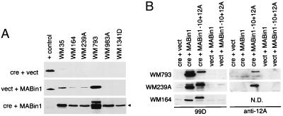
Figure 5.
Bin1 expression from cre-inducible adenoviral vectors. (A) Bin1 expression. The melanoma cells indicated were infected with Ad-cre + Ad-vect (Top), Ad-vect + Ad-MABin1 (Middle), or Ad-cre + Ad-MABin1 (Bottom). Cell lysates were prepared 48 hr later and subjected to Western blotting with Bin1 antibody 99D. The positive control (+ control) for expression is an extract from murine C2C12 cells, which robustly express the −10+13 splice isoform (3). The exposure times were short to identify only exogenous levels of Bin1 expression (exposure in Bottom was shortest). (B) Bin1–10+12A expression. Cells indicated were infected and analyzed as above, except that Ad-MABin1−10+12A was used in addition to Ad-MABin1 and parallel blots were processed with 99D or anti-12A antibodies. N.D., not done.

