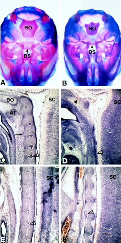
Figure 4.
Analysis of the chondrocranium (A and B at E18.5) and notochord (C–F at E14.5) Ventral views of the skull of wild type (A) and mutant (B) show a decrease in size of the basioccipital bone (BO) and posterior basisphenoid bone (BS). C–F show sagittal sections of wild-type (C and E) and mutant (D and F) embryos near the midline of the axial skeleton through the notochord region. C and D are sections from the cervical region. The characteristic notochordal swellings are shown in C (open arrowhead indicates characteristic swelling). The thin mutant notochord is indicated by the open arrowhead in D. The condensing cells of the anterior located basioccipital bone (in C, designated BO) are missing in the mutant (region indicted by filled arrowhead in D). More posteriorly, the periodic notochordal swellings in wild type (open arrowhead in E) are abnormal in the mutant (F). AT, anterior arch of the atlas; SC, spinal cord; BO, basioccipital; BS, posterior basisphenoid.

