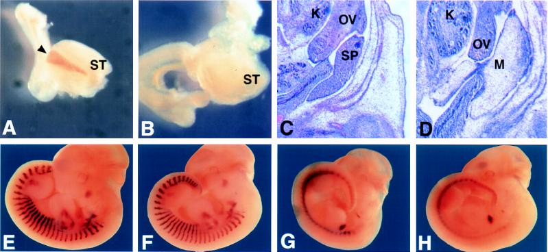
Figure 5.
Analysis of the spleen and expression of Hox11, myogenin, and Pax1. Expression of Hox11 at E12 was examined in stomach and surrounding tissue from dissected embryos. The spleen of wild-type (A) embryos is observed as a brown stripe next to the stomach (black arrowhead), which is missing in Bapx1−/− (B). Histological examination in transverse sections shows densely packed cells of the spleen in wild type (in C, designated SP), which is not observed in a comparable region of mesentery in the mutant (D). Wild type (E and F) and mutant (G and H) show no differences in the expression of myogenin (lateral stripes in E and F) or Pax1 (lateral periodic spots in G and H). ST, stomach; K, kidney; OV, ovary; SP, spleen; M, mesentary.

