
Fig. 1.
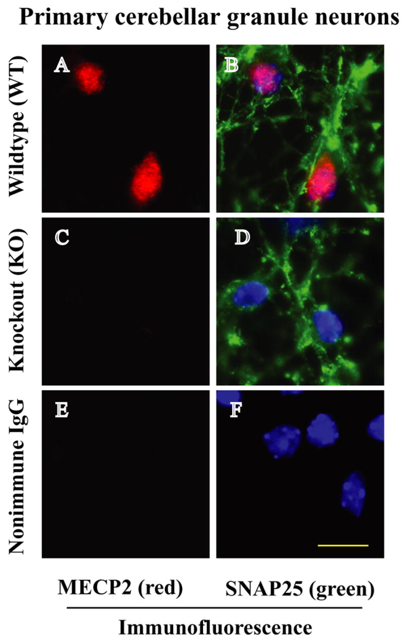
Images of immunofluorescence staining of MeCP2 in WT and MeCP2 KO cerebellar granule neurons. A) MeCP2 is expressed (red staining) in WT cells. B and D) SNAP25 (green staining) is a neuron specific marker that stains the cerebellar granule neurons. Moreover, the same cells were stained with DAPI, a dye that binds nucleic acid and labels cell nuclei. Digitized individual and overlapped images of MeCP2, SNAP25 and DAPI in B indicate that MeCP2 is localized in the nucleus of cerebellar granule neurons. Panel (C) shows complete absence of MeCP2 proteins in MeCP2−/− cerebellar granule neurons (D). Non-immune IgG staining shows no evidence of MeCP2 or SNAP25 (E, F). Scale bar 10 μm.