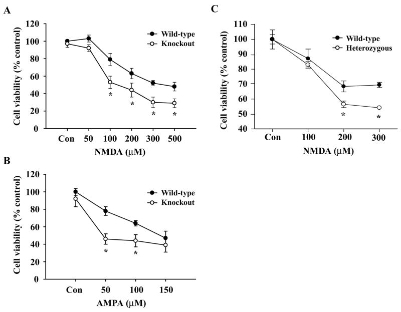
Fig. 2.
Effects of NMDA and AMPA-induced excitotoxicity on WT and MeCP2−/−primary cerebellar granule neurons. CGC cultures (DIV 10) were exposed to increasing concentrations of NMDA (A, C) and AMPA (B) for ~20 h at 37°C. Quantification of neuronal viability by MTT reduction colorimetric assays revealed that 200 μM of NMDA (A) and 100 μM of AMPA (B) resulted in ~ 50% cell death in MeCP2−/− cells (p<0.01) compared to WT controls. MeCP2 heterozygous cells also had a significantly higher percentage of cell death than WT controls. Values are shown as mean ± SEM of % cell viability relative to controls (100%) from 6 independent observations (n=6). *p<0.01

