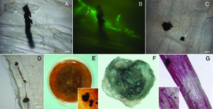
FIG. 4.
MS of Colletotrichum graminicola. Roots were excised from maize seeds grown in sterile vermiculite inoculated with agar plugs colonized with vegetative mycelium of C. graminicola expressing GFP (A to D). MS of C. graminicola M1.001-BH-gfp were produced in vitro (E to G). Photographs were obtained by bright-field microscopy (A, C, and D), by fluorescence microscopy (B), or under a stereomicroscope (E to G). (A and B) MS of C. graminicola attached to hyphae (arrow) on the maize root surface at 10 dpi, viewed with bright-field (A) and fluorescent (B) illumination. (C) MS of C. graminicola (arrow) on the maize root; 7 dpi. Notice the variation in MS size and shape. (D) Many MS on the root; 20 dpi. (E) MS produced in vitro. (F) Cleaned MS produced in vitro. Masses of salmon-orange conidia (arrow) can be observed on the surface of the MS. (G) Germination of MS on roots. Melanized hyphae and hyphopodia (arrow, inset) can be observed colonizing the root surface; 7 dpi. Bars = 10 μm (A, B, and C) and 20 μm (D).

