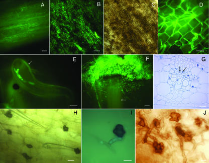
FIG. 5.
Colletotrichum graminicola spreads systemically from infected roots to aerial plant parts. Plants were from maize seeds grown in a sterile vermiculite system and inoculated with agar plugs of M1.001-BH-gfp mycelium. Photographs were obtained using bright-field microscopy (C, G, and H), using fluorescence microscopy (A to B, D to F, and I), or under a stereomicroscope (J). (A) Adventitious root of maize systemically colonized by C. graminicola; 28 dpi. (B and C) Senescent coleoptile colonized by C. graminicola; 28 dpi. Viewed with fluorescence (B) and bright-field (C) illumination. (D) Colonization of the third leaf sheath from a root-infected plant; 28 dpi. (E) Hyphae growing inside of a leaf trichome (arrow). (F) C. graminicola growing from individual vascular bundles (arrow) of a stem cut in transverse section and cultured on isolation medium. (G) Five-micrometer cross section of a maize leaf from a root-inoculated plant; 28 dpi. Hyphae are indicated by arrows. (H) Lobed hyphopodia on an infected, senescent leaf sheath; 28 dpi. (I) Hyphopodia begin to accumulate melanin, though GFP fluorescence within the hyphae is still observed in the hypha leading to the hyphopodium. (J) Lobed hyphopodia produced in liquid culture; 3 dpi. Bars = 100 μm (A and F), 20 μm (B, C, and G), and 10 μm (D, E, H, I, and J).

