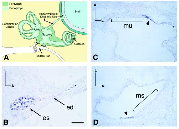
Figure 2.
RNA in situ hybridization of Pds in noncochlear regions of the mouse inner ear. (A) Anatomy of the fully developed mammalian inner ear (adapted from ref. 46). (B–D) Transverse sections (12 μm) through the inner ear of a P5 mouse were obtained and hybridized with a Pds-specific riboprobe. Note the detection of Pds expression (indicated by arrowheads) throughout the endolymphatic duct and sac (B) and in nonsensory regions of the utricle (C) and saccule (D), adjacent to the maculae in both of the latter cases. es, endolymphatic sac; ed, endolymphatic duct; mu, macula utriculi; ms, macula sacculi. Orientations: A, anterior; L, lateral. (Bar = 100 μm.)

