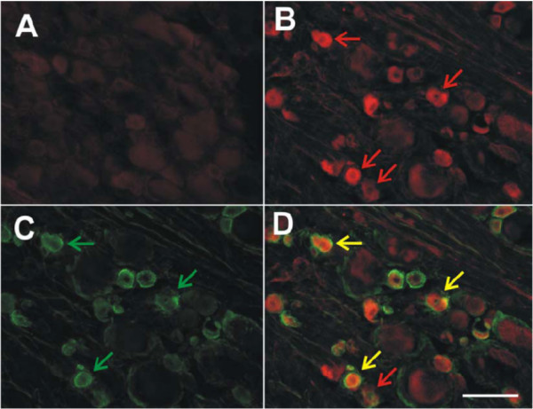
Figure 6.
Colocalization of MCP-1 immunoreactivity (ir) and isolectin B4(IB4)-binding in the lumbar DRG ipsilateral to LPC-induced demyelination injury at POD7. IB4-binding in rat DRG neurons distinguishes C-fiber nociceptors. (A) Naïve rat lumbar DRG were completely negative for MCP-1ir. (B) Lumbar DRG ipsilateral to LPC-induced sciatic nerve injury exhibited numerous small diameter neurons that are MCP-1 positive at POD7 (red arrows). (C) Numerous small IB4-binding presumptive nociceptors are present in the same DRG tissue section (green arrows). (D) Merging panels B and C demonstrates the extent of colocalization present in lumbar DRG tissue section (yellow arrows). Note not all MCP-1ir neurons were positive for IB4 at POD7 (red arrow). Scale bar is 100 μm (A, B, C and D).

