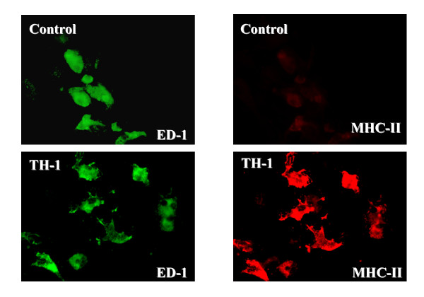
Figure 1.
Effect of Th1 cytokines on microglia. CNS mixed glial cultures were incubated with Th1 cytokines or additional culture medium for 4 days. Cultures were fixed with 4% paraformaldehyde for 10 minutes, washed and then incubated with mouse anti-ED-1 (IgM) and mouse anti-rat MHC class II (IgG) followed by Alexa 488-conjugated goat anti-mouse IgM and Cy 3-conjugated donkey anti-mouse IgG. Cultures were examined for indirect immunofluoresence employing a Leitz Orthoplan 2 fluorescent microscope. The Th1 treated microglia (ED-1+ cells) have a different appearance when compared to those incubated with additional medium (control). Control microglia do not express MHC class II whereas Th1 treated microglia strongly express MHC class II molecules.

