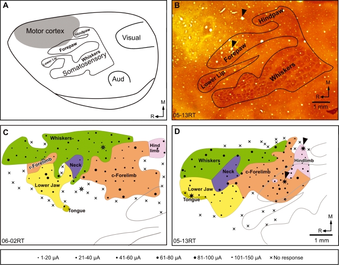
Fig. 2.
(A) An outline diagram of the dorsolateral view of the cerebral hemisphere of a rat brain showing the location of the motor cortex and its relation to major sensory areas. Body parts in the somatosensory cortex S1 are marked; Aud., primary auditory cortex. (B) A photomontage showing the somatosensory isomorph revealed in sections of the flattened cortex stained for cytochrome oxidase activity, for rat 05–13RT. The location and details of the motor cortical map are described in relation to such somatosensory isomorphs. Arrowheads point to the microlesions used to align it with the motor map shown in (D). (C and D) Organization of the motor cortex in two rats mapped under light anaesthesia. Note the large medial representation of the whiskers and the smaller neck region. The rostral (r) and the caudal (c) forelimb regions are marked. (D) In rat 05–13RT the rostral forelimb was not seen, probably because of low mapping density in the region due to an overlying blood vessel. The grey outlines show portions of the somatosensory isomorph (see A and B). The bottom panel shows the key to the symbols used in C and D. The stars mark the locations of microlesions used to align topographic map to the somatosensory isomorph. R, rostral; M, medial. The scale bar corresponds to the distance on the brain measured during recordings. Scale bar and the orientation arrows shown in D are also valid for C

