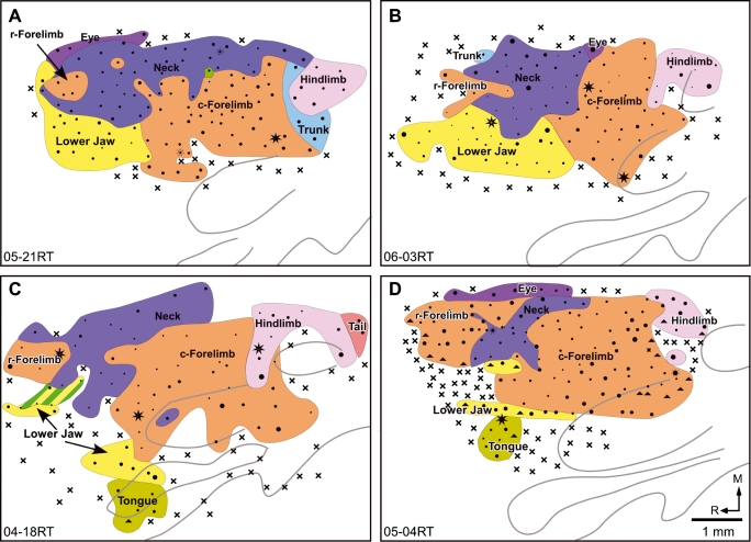
Fig. 3.
Organization of the motor cortex in four rats (A–D) mapped under deep anaesthesia. Note the absence of the whisker region except for a single point in (A) and two points overlapping the jaw region in (C) The neck representation is enlarged as compared to rats mapped under light anaesthesia (cf. Fig. 2C and D). Other conventions as for Fig. 2.

