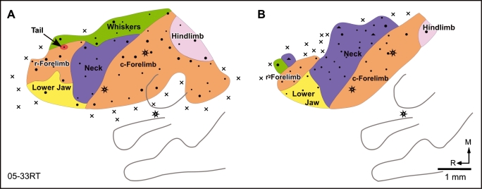
Fig. 4.
Organization of the motor cortex in a rat mapped (A) first under light and (B) then under deep anaesthesia. Under deep anaesthesia the whisker movements could not be evoked except for one point in the rostralmost region. The neck is greatly expanded, particularly into the caudal whisker region. The caudolateral region of the motor cortex could not be mapped under deep anaesthesia because the cortex became unresponsive due to the length of the experiment. Other conventions as for Fig. 2.

