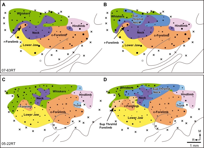
Fig. 5.
Maps of the motor cortex of two rats at (A and C) threshold currents, and (B and D) when movements at higher currents were considered. Note that at suprathreshold currents the neck movement region expanded into the caudal part of the whisker region. In rat 05–22RT an expansion of the rostral forelimb region is also seen. The whisker movements evoked along with the expanded neck or the rostral forelimb regions are not illustrated in B and D. Sup Thrshld, suprathreshold. Other conventions as for Fig. 2.

