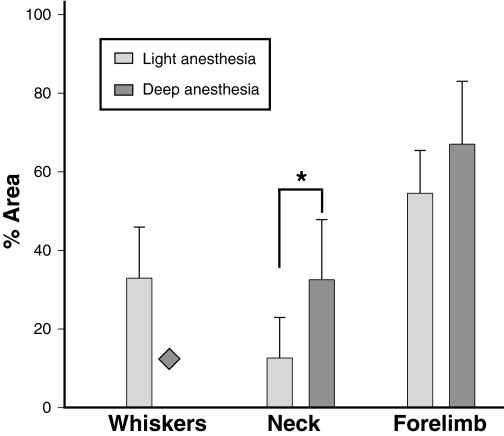
Fig. 8.
Areas of representation of the whisker, neck and forelimb regions in the motor cortex as a percentage of total area occupied by these representations when the rats were mapped under light and deep anaesthesia. There was no whisker movement under deep anaesthesia ( ). Both the neck and forelimb areas were larger under deep anaesthesia; however, only the areas of the neck representations were significantly different (*P = 0.011; unpaired t-test).

