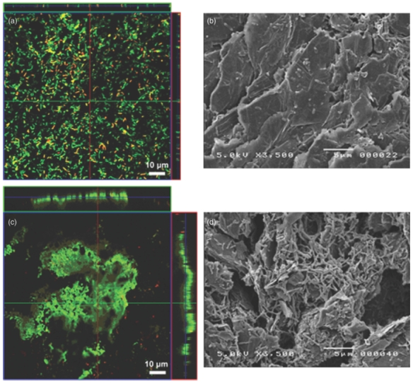
Fig. 3.

Confocal (a, c) and SEM (b, d) pictures of the biofilm growing on anodes of fuel cells inoculated with Shewanella oneidensis MR-1. Fuel cells were fed with FW medium supplemented with lactate and (a, b) amino acids; (c, d) yeast extract. Anodes for CLSM examination were directly stained with the BacLight Live/Dead kit (Korber et al., 1996). Live cells appear green and dead cells red.
