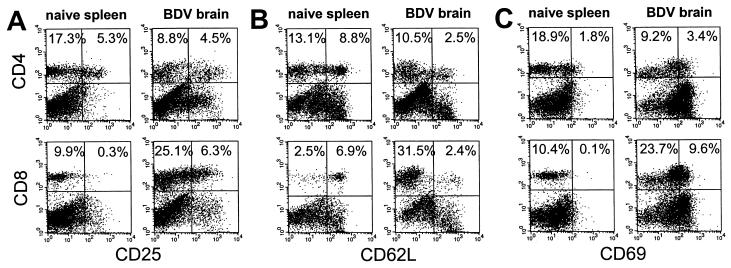
Figure 1.
Expression of activation markers on brain-derived CD8+ T cells. Neonatally infected MRL mice were sacrificed at the peak of neurological disease, and brain lymphocytes were isolated. Uninfected age-matched MRL mice served as donors for spleen lymphocytes. Lymphocytes were stained with anti-CD8-FITC or anti-CD4-FITC and biotinylated mAb specific for CD25 (IL-2 receptor) (A), CD62L (l-selectin) (B), or CD69 (very early activation antigen) (C) followed by streptavidin-R-PE detection. Three independent experiments were performed and dot plots are shown from one representative experiment. (Left) Analysis of spleen lymphocytes from a naive mouse. (Right) Brain-derived lymphocytes. Percentages of CD4+ and CD8+ T cells positive for the indicated marker are given in the upper quadrants.

