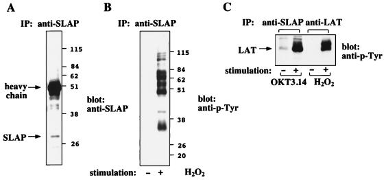
Figure 2.
SLAP is associated with several tyrosyl-phosphorylated proteins in Jurkat T cells. (A) Jurkat T cell lysate (from 2 × 10⁷ cells) was incubated with affinity-purified anti-SLAP antibody and protein-A-Sepharose. Immunoprecipitated proteins were separated on 10% SDS/PAGE and blotted with anti-SLAP antibody. The position of SLAP at about 34 kDa is indicated. (B) Jurkat T-cells (2 × 10⁷) were stimulated with 5 mM H2O2 at 37°C for 3 min. Stimulated and nonstimulated cells lysates were immunoprecipitated with affinity-purified anti-SLAP antibody. Immunoprecipitated proteins were separated on 10% SDS/PAGE and blotted with anti-p-Tyr antibody. (C) Jurkat T-cells (5 × 10⁷) were incubated with monoclonal anti-CD3 antibody at 4°C for 10 min. Cells were washed with PBS and incubated with 10-fold sheep anti-mouse antibody conjugated to Dynabeads at 4°C for 10 min. The cell-beads mixture was incubated at 37°C for 3 min and lysed with lysis buffer containing 1% NP-40. Dynabeads were removed from the lysate by centrifugation. Cell lysates were incubated with protein A-Sepharose for 1 hr before the supernatant was immunoprecipitated with affinity-purified anti-SLAP antibody. Protein complexes were separated by 10% SDS/PAGE and blotted with anti-p-Tyr. The anti-LAT immunoprecipitation of H2O2-stimulated and nonstimulated lysates were run as a marker for LAT protein.

