Figure 3.
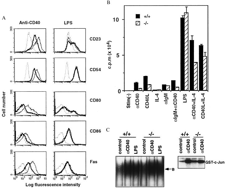
CD40-mediated B cell activation in vitro. (A) Up-regulation of CD23, CD54, CD80, CD86, and Fas by anti-CD40 or LPS stimulation. Purified B cells from traf5+/+ (thin line) and traf5−/− (thick line) were stimulated with anti-CD40 mAb or LPS or were unstimulated (dotted line). After 48 hr, the cells were stained with FITC-labeled anti-CD23, anti-CD54, anti-CD80, anti-CD86, or anti-Fas mAb. Data represent one of three independent experiments with similar results. (B) Proliferative responses. Purified B cells isolated from traf5+/+ (solid bars) and traf5−/− (hatched bars) mice were stimulated for 48 hr with anti-CD40 mAb, CD40L-CD8 chimeric protein (CD40L), IL-4, anti-IgM Ab, or LPS. Proliferation was assessed by [3H]thymidine incorporation during the last 8 hr. Data are shown as the mean cpm ± SEM from triplicate samples and represent one of three independent experiments with similar results. (C) Splenocytes (for EMSA) or purified B cells (for in vitro kinase assay) isolated from traf5+/+ and traf5−/− mice were stimulated with anti-CD40 mAb for 15 min or LPS for 30 min. EMSA for NF-κB activation (Left) and in vitro kinase assay for JNK/SAPK activation (Right) were performed as described in Materials and Methods. B, oligonucleotide probe bound to NF-κB complexes.

