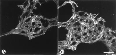
Figure 10.
Confocal micrographs of pulmonary vessels stained by injection of rhodamine-labeled Ricin lectin. (A) Alveolar capillaries in mouse given rat IgG. Luminal surface of endothelial cells stains uniformly faint whereas adherent leukocytes stain more brightly (arrowhead). (B) Alveolar capillaries in mouse given BV13 (50 μg, 4 hours). Bright focal patches of staining (arrows) indicate exposed basement membrane. (Bar = 25 μm.)

