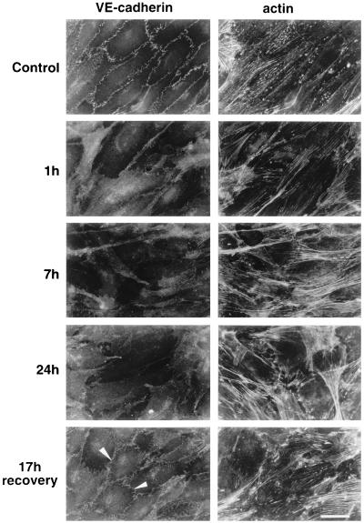
Figure 2.
Effect of BV13 on VE-cadherin organization in endothelial cells. BV13 (50 μg/ml) was added to cultured endothelial monolayers (1G11). VE-cadherin staining was strongly reduced at junctions within 1 hour, and this effect lasted up to 24 hours of incubation with the mAb. When BV13 was removed after 7 hours of incubation and the cells were cultured for additional 17 hours, a partial recovery of VE-cadherin at junctions was detected (white arrowheads). Actin staining shows that reduction of VE-cadherin staining from junctions was not accompanied by cell retraction. Comparable results were obtained when, after fixation of the cells, VE-cadherin was detected by using either mAb BV14 or a VE-cadherin rabbit polyclonal antiserum (data not shown). (Bar = 20 μm.)

