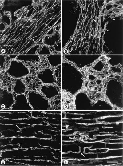
Figure 6.
Endothelial cell junctions stained by i.v. injection of VE-cadherin antibody. Blood vessels were labeled by i.v. injection of BV13 and ex vivo incubation in Cy3-labeled secondary antibody. Images of vessels in thick tissue sections were obtained by fluorescence confocal microscopy. Shown are intrapulmonary segments of pulmonary arterioles at 20 minutes (A) or 2 hours (B) after i.v. injection of BV13. In the large arterioles and venules, antibody bound continuously along endothelial cell–cell junctions at 20 minutes and 2 hours (arrows in A and B). In small pulmonary vessels near arterioles, antibody also bound in sharp, continuous lines along endothelial cell junctions at 20 minutes (arrowheads in A), but, in contrast, binding was discontinuous and diffuse at 2 hours (arrowheads in B). Similarly, in alveolar capillaries, antibody bound in sharp, continuous lines at 20 minutes (arrow in C), but binding was discontinuous and diffuse at 2 hours (arrow in D). In capillaries of cardiac muscle, antibody bound in sharp, continuous lines at 20 minutes (arrow in E) but was discontinuous and diffuse at 2 hours (arrow in F). (Bar = 25 μm.)

