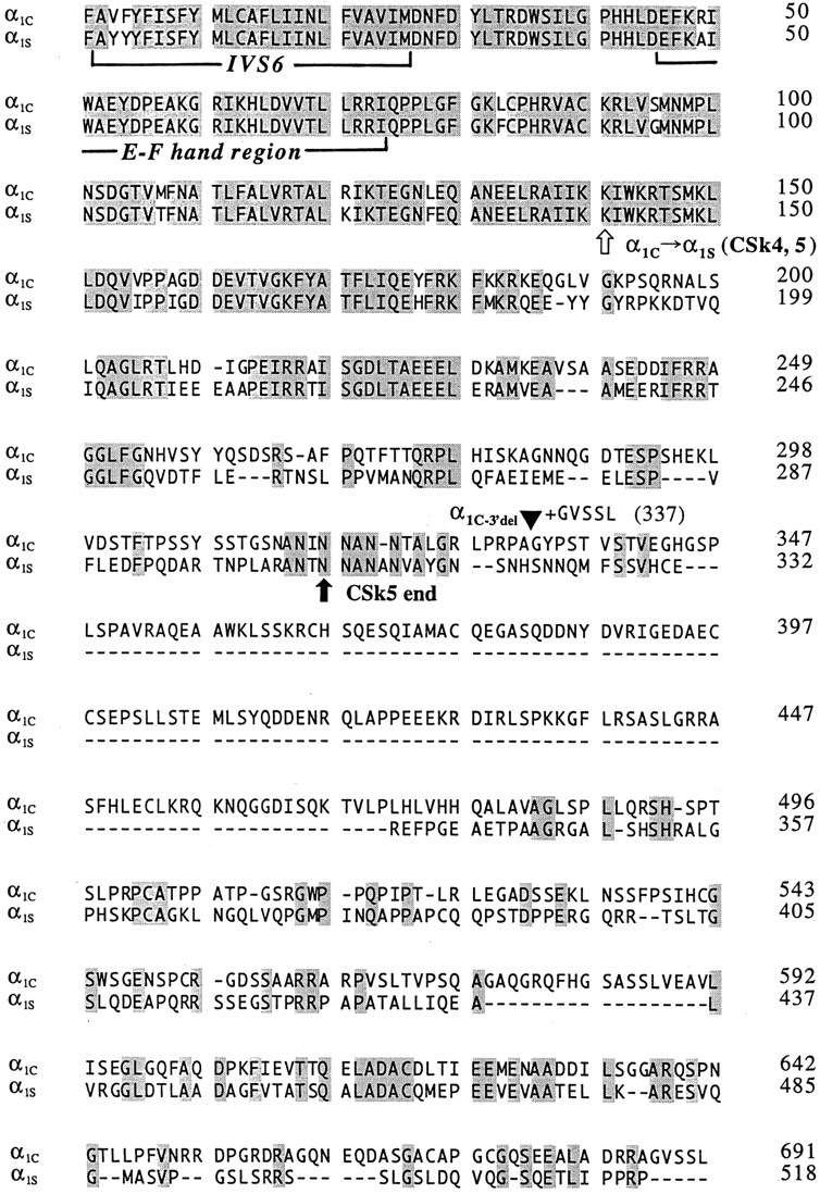
Figure 7.

Sequence alignment of the COOH-terminal regions of the rabbit cardiac α1C (Mikami et al., 1989) and rabbit skeletal muscle α1S subunits (Tanabe et al., 1987). The sequences begin just before the last predicted transmembrane segment (S6) of domain IV (first bracket). The putative EF hand motif is indicated (second bracket), as is the junctional site within CSk4 and CSk5 where the sequence changes from α1C to α1S (white arrow). The termination of mutant α1C−3′del is indicated by a filled triangle; this construct retains the final five residues (GVSSL) of the wild-type COOH terminus. The termination point of chimera CSk5 is indicated (black arrow). Shaded amino acids are identical between α1C and α1S; dashes indicate gaps in the alignment.
