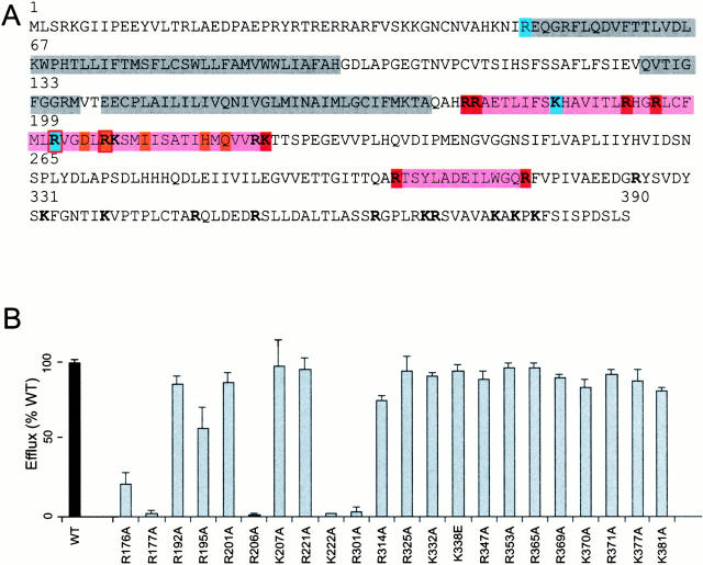
Figure 1.
Alanine scanning mutagenesis of COOH-terminal basic residues. (A) Primary sequence of Kir6.2, highlighted to show transmembrane M1 and M2 domains together with the P-loop (gray), and regions that enclose PIP2-sensitive residues (pink). All residues that were mutated in this study are indicated (bold), including those that alter PIP2 response in this study (red) or ATP sensitivity (blue). Also indicated are the PIP2-sensitive residues identified in Kir2.1/Kir3.1 by Zhang et al. 1999(orange). (B) 86Rb efflux in 40 min from COSm6 cells cotransfected with mutant Kir6.2 subunits (as indicated) + SUR1, expressed as a percentage of the efflux from cotransfected wild-type Kir6.2 + SUR1 subunits in parallel transfections (mean ± SEM, n = 3 in each case). See materials and methods for details.

