Figure 1.
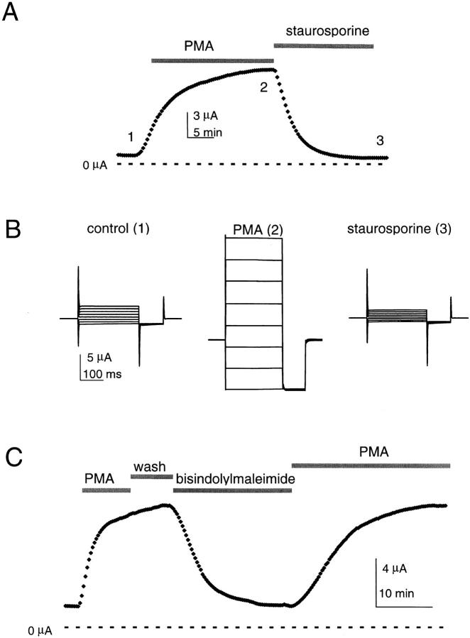
Modulators of PKC determine KCNKØ current magnitude. Macroscopic KCNKØ channels currents measured by two-electrode voltage clamp in 20 mM potassium solution with or without 50 nM PMA or 2 μM staurosporine (see materials and methods). (A) Currents measured during the application of PMA and then staurosporine; the oocyte was held at −40 mV and stepped to 25 mV for 250 ms with a 20-s interpulse interval. The response to PMA varied among batches of oocytes from 3–11-fold; this appeared to reflect different levels of prior activation as a consistent 10 ± 1-fold increase in currents was observed when PMA and staurosporine treatments were compared (mean ± SEM, n = 10). Zero current is indicated. (B) Raw current traces for the oocyte in A at various times as follows: (1) control solution; (2) after 20 min in PMA; and (3) after 20 min in staurosporine. The cell was held at −80 mV and studied at these times by 250-ms steps from−150 to 60 mV in 30-mV increments, and then studied for 75 ms at −150 mV with a 2-s interpulse interval. (C) Currents measured at 25 mV by the protocol in A during 10-min activation by 50 nM PMA, 6-min wash with control solution, 2-min inhibition by 4 μM bisindolylmaleimide I, and then a 20-min reactivation by 50 nM PMA. Zero current is indicated.

