Figure 4.
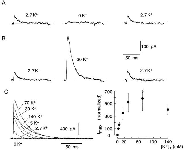
Modulation by extracellular K+ of E418Q channels. (A and B) Reversible modification of the amplitude of K+ currents recorded in control conditions (2.7 mM K+) and during exposure to low (0 mM) or high (30 mM) extracellular [K+]. Internal [K+] was 130 mM. In all cases, 100-ms pulses were applied to +20 mV. (C, left) K+ currents recorded from a cell with a pulse to +20 mV during exposure to various extracellular [K+]. (Right) Changes of peak K+ current amplitude at +20 mV (ordinate) as a function of extracellular [K+] (abscissa). Data points are the mean and the vertical bars are the standard deviation of measurements done in eight cells.

