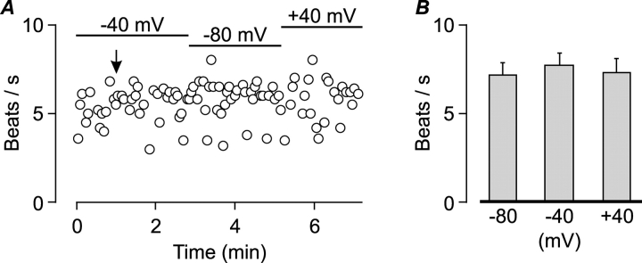
Figure 7.
Spontaneous beating is not modulated by the membrane potential. (A) An example of CBF measurement from a single isolated cell voltage clamped at either −40, −80, or 40 mV as indicated by bars. The arrow indicates the time of whole-cell formation. Standard extracellular and pipette solutions. (B) Mean (±SEM) CBF of seven ciliated cells voltage-clamped at −80, −40, and 40 mV from experiments of the type shown in A. There was no significant difference between groups (Student's t test).

