Figure 1.
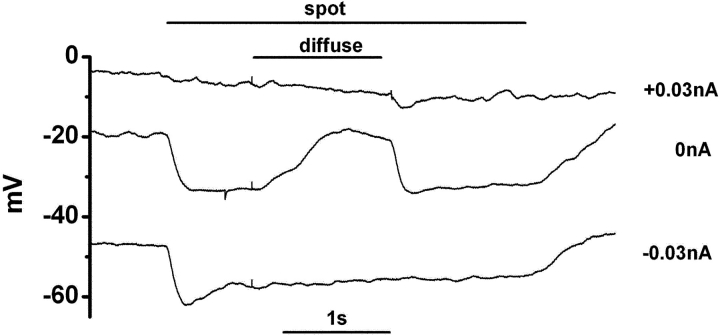
The response of a newt cone photoreceptor recorded in the current-clamp mode The outer segment of the cone was illuminated by a spot (diameter, 30 μm; duration, 3,380 ms; timing indicated by the top horizontal line). A diffuse light (diameter, 4,000 μm; duration, 1,250 ms) was superimposed on the spot as indicated by the shorter horizontal line. The retinal slice was superfused with control Ringer's solution buffered with bicarbonate and containing 100 μM picrotoxin. Under control condition (when no current was injected from the recording pipette: 0 nA), illumination with the spot evoked hyperpolarization, and the surround illumination evoked depolarization in the cone. Both hyperpolarization and depolarization of the cone induced by current injection (−0.03 and +0.03 nA) from the recording pipette abolished the surround response. The vertical scale on the left indicates the absolute membrane voltage. Recovery at the spot offset was slow (1 s), probably due to blockade of the calcium feedback to the phototransduction cascade in the cones (Lamb et al., 1986; Nakatani and Yau, 1988), because the intracellular Ca2+ level was maintained at a low level due to the addition of 20 mM BAPTA in the pipette solution. Lowering the BAPTA concentration in the pipette solution (5 mM) accelerated the recovery (0.5 s; unpublished data).

