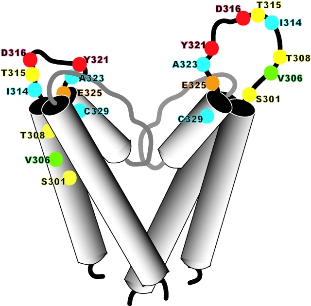
Figure 11.
Contrasting models of the CNG channel pore turret. The structure of the left-hand subunit is based on the KcsA structure with a shorter pore turret, and the model that we propose with a longer pore turret is shown on the left. Amino acid residues that were mutated in the current study are indicated as balls on the ribbon backbone. The strength of interaction is shown by a color scale, ranging from blue (undetectable) to yellow (weak) to orange (moderate) to red (strong). The green residue indicates the position homologous to that of a known glycosylation site in CNGA1.

