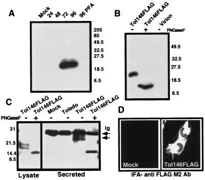
Figure 2.
Expression of FLAG-tagged vCXC-1 by Tol146FLAG. (A) Detection of FLAG-tagged vCXC-1 in cell lysates by immunoblot with anti-FLAG M2 Ab. Whole-cell lysates were harvested from uninfected (Mock) or Tol146FLAG-infected cells at 24, 48, 72, and 96 hpi. Cell lysates also were collected at 96 hpi from Tol146 FLAG-infected cells cultured in the presence of 660 μM phosphonoformic acid (96 PFA). (B) Structural analysis of FLAG-tagged vCXC-1. Infected cell lysates and virions, enriched by sucrose gradient, were harvested at 96 hpi and treated with PNGase F as indicated (+). The proteins were immunoblotted with anti-FLAG M2 Ab [Coomassie staining confirmed the composition of the virion preparation: viral (pp65) but not cellular (β-actin) antigens were detected by immunoblotting.] (C) Immunoprecipitation of FLAG-tagged vCXC-1 from medium (Secreted) and cell lysates. Protein was harvested at 96 hpi from mock-infected, Toledo, and Tol146FLAG-infected fibroblasts, treated with PNGase F as indicated (+), and precipitated with anti-FLAG M2 Ab. The arrow labeled “Ig” marks a nonspecific reaction with Ig light chain. The arrow below denotes the position of 25,000- to 27,000-MW secreted form of FLAG-tagged vCXC-1. (D) Mock- and Tol146FLAG-infected fibroblasts were stained with anti-FLAG M2 Ab and goat anti-mouse FITC at 96 hpi. Toledo-infected and antibody controls were negative.

