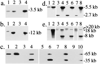
Figure 3.
Southern blot analysis of S. typhimurium M4A36; aphT-Φ. (a) Southern blot analysis of EcoRV-digested chromosomal DNA using probe I (see Fig. 2). (b) Southern blot analysis of HindIII-digested chromosomal DNA using probe IV. Bands in lanes 2 and 4 were shifted because of the presence of the aphT cassette. DNA was from 1, 3351/78DT204;E+; 2, M1063351/78;aphT-Φ; 3, A36DT36;E−; and 4, M4A36;aphT-Φ. (c) Chromosomal DNA was digested with XbaI and analyzed by PFGE and Southern blot hybridization by using probe VI (see Fig. 2). The S. typhimurium strains were: 1, 3351/78DT204;E+; 2, M1063351/78;aphT-Φ; 3, A36DT36;E−; 4, M4A36;aphT-Φ; 5, 3805/96DT186;E−; 6, M63805/96;aphT-Φ; 7, 3739/96DT193;E−; 8, M93739/96;aphT-Φ; 9, 2138/96DT120;E−; and 10, M102138/96;aphT-Φ. (d and e) Chromosomal DNA from strains 1, 3351/78DT204;E+; 2, M1063351/78;aphT-Φ; 3, A36DT36;E−; 4, M4A36;aphT-Φ; 5, 3805/96DT186;E−; 6, M63805/96;aphT-Φ; 7, 2138/96DT120;E−; 8, M102138/96;aphT-Φ was digested with EcoRV (d) or HindIII (e) and analyzed by using probe VIII (see Fig. 2).

