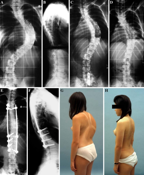
Fig. 3.
Subject 4 preoperative ap and lateral radiographs showing right convex thoracic scoliosis of 84° (a, b). The preoperative ap traction radiograph shows scoliosis correction to 68° (c) and traction films after anterior release from Th6–L1 shows correction to 55° (d). Postoperative pictures showing correction to 23° (e, f). Clinical photographs showing the preoperative deformity with a significant rib hump (g) and the postoperative result (h)

