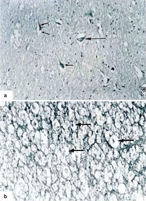
Fig. 3.
Three hours of ischemia. A (100×) Neuronal degeneration and death (small arrows), neurons and vessels with clear halos surrounding the nucleus (big arrow). Asterisk shows a damaged vessel. B (400×) White matter with neuronal degeneration and chromatolysis (arrows). Hematoxylin and eosin staining

