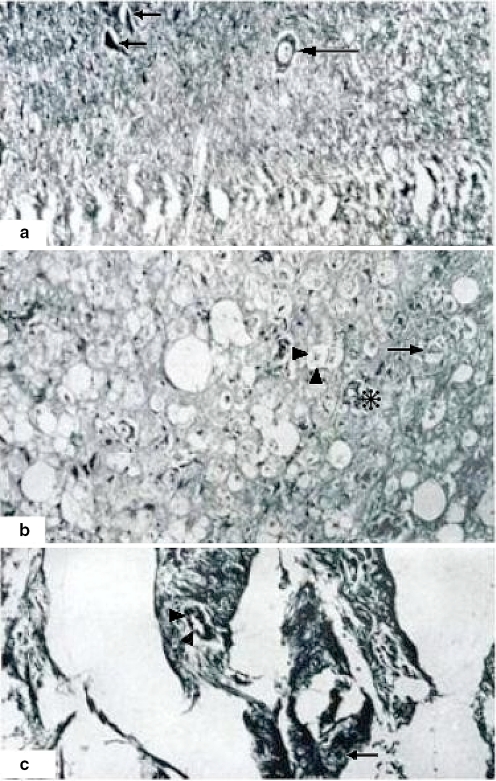
Fig. 4.
Three hours of ischemia with extensive chromatolysis. A (100×) Neuronal degeneration and chromatolysis (small arrows) and not completely damaged (just a clear halo surrounding it) neurons (big arrow). B (400×) White matter with altered cords. Some of the nerves show necrosis and severe dissolution (arrowheads). However, in other areas nerves are intact (big arrows). Asterisk shows a small normal vessel. C Scare zone with fibrosis and general severe damage and total loss of cytoarchitecture. There is also a degenerated neuron (small arrow) and a damaged vessel (arrowheads). Hematoxylin and eosin staining

