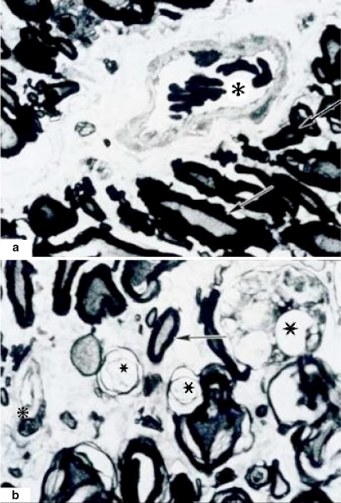
Fig. 6.
Semifine coronal sections. Myelin dissociation at 3 h of ischemia. A (800×) Proximal section with a segment of preserved fascicules. Some axons with discrete myelin dissociation (arrows) are observed. There is a normal vessel (asterisk). B (800×) Distal section. Almost total degeneration of axons with elongation and dissociation of myelin and multiple axonal remainders (stars). A vessel with hypertrophic endothelium is showed (small asterisk). There is a normal axon (arrow) surrounded by a necrotic area. Tissue samples in semifine cross sections were embedded in epoxy resin (poly/bed) 1 μm in width. Toluidine blue method staining

