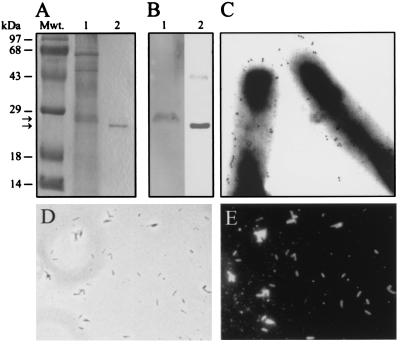
Figure 3.
ML-LBP21 is a major cell wall-associated and surface protein of M. leprae. (A) Coommassie blue-stained SDS/polyacrylamide gel of M. leprae cell wall fraction (lane 1) and rML-LBP21 (lane 2). Molecular mass markers (Mwt) in kDa are indicated above the left lane. (B) Corresponding immunoblot of M. leprae cell wall fraction (lane 1) and rML-LBP21 (lane 2) was labeled with mAb raised against rML-LBP21. mAb-reactive single 28-kDa protein band (B, lane 1) is a major protein in the cell-wall fraction (A, lane 1) that corresponds to rML-LBP21 (B, lane 2). (C) Immunoelectron microscopy on intact M. leprae with mAb against rML-LBP21. Colloidal gold particles represent the localization of ML-LBP21 on the surface of M. leprae. (D and E) Indirect immunofluorescence staining of intact M. leprae with mAb to rML-LBP21 demonstrating the intense labeling in all bacilli in the field (E). Corresponding phase contrast image is shown in D.

