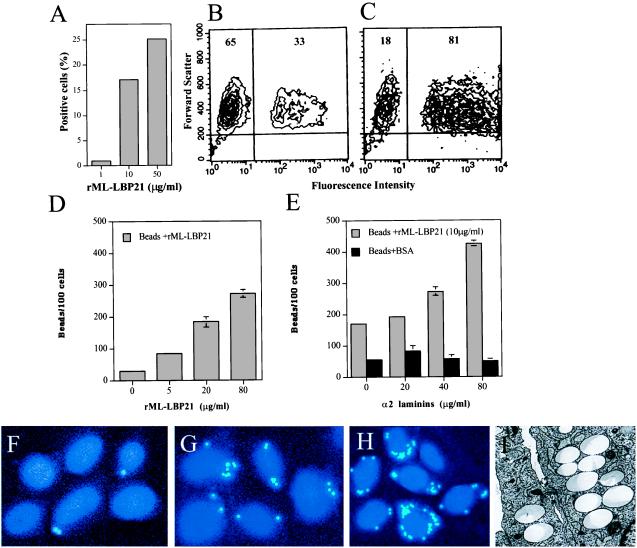
Figure 5.
Role of rML-LBP21 in Schwann cell adherence and invasion. (A) Adherence of rML-LBP21-coated fluoresceinated beads to primary Schwann cells in suspension in the absence of α2 laminins as analyzed by flow cytometry. A representative example showing the percentage of positive cells carrying fluoresceinated beads coated with increasing concentration of rML-LBP21. (B and C) Representative FACS plots of Schwann cell-bound rML-LBP21-coated beads in the absence (B) and the presence of α2 laminins (C). Numbers indicate the percentage of total Schwann cells within the indicated gate. (D–I) Schwann cell invasion of rML-LBP21-coated beads in the absence and the presence of exogenous α2 laminins as determined by fluorescence and electron microscopy. (D) Quantification of Schwann cell invasion of beads coated with increasing concentration of rML-LBP21 alone as determined by fluorescence microscopy. (E) Schwann cell invasion of rML-LBP21- and BSA-coated beads in the presence of increasing concentration of exogenous α2 laminins. Fluorescence microscopic images of Schwann cell-invaded beads coated with BSA (F) rML-LBP21 (G) and rML-LBP21 + α2 laminins (H). Intracellular beads and Schwann cell nuclei are shown in light blue (dots) and blue, respectively. (I) Electron micrograph of Schwann cells showing the intracellular location of beads coated with rML-LBP21 + α2 laminins.

