
Fig. 5.
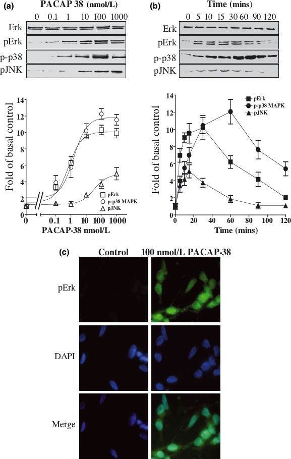
PACAP-38-mediated activation of ERK, p38 MAP kinase and JNK. SH-SY5Y cells were cultured in low serum medium for 24 h and incubated with increasing concentrations of PACAP-38 for 15 min (a) or with 100 nmol/L PACAP-38 for up to 2 h (b). Western blot analysis of whole cell extracts was carried out using antibodies specific for ERK, pERK, p-p38 MAP kinase and pJNK. The increase in phosphorylation levels was determined by densitometry as described in Materials and methods and is graphically represented in the lower section of the figure. Standard error bars are shown (n = 5). (c) The cells were incubated with a pERK-specific antibody and its respective fluorescent dye-conjugated secondary antibody (green) following treatment with 100 nmol/L PACAP-38 for 30 min. Cells were also stained with DAPI, the DNA-binding fluorescent stain (blue). Each micrograph is representative of three independent experiments.