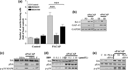
Fig. 6.
The effect of ERK and p38 MAP kinase on PACAP-induced differentiation of SH-SY5Y cells. (a–b) SH-SY5Y cells were cultured in low serum medium for 24 h and incubated with 10 μmol/L PD98059 (PD) or 10 μmol/L SB203580 (SB) for 45 min before treatment with 100 nmol/L PACAP-38 or low serum medium as control for 4 days. Treatments were replaced after the second day. (a) The fold increase in neurite-bearing cells was determined as described in Materials and methods. Standard error bars are shown (n = 3) and ANOVA was carried out to identify significant differences between treatments (***p < 0.001). (b) Western blot analysis of whole cell extracts using antibodies specific for Bcl-2 and GAP-43 was carried out on day 4. Protein loading was determined using an antibody specific for GAPDH. (c-e) Western blot analysis of whole cell extracts was carried out using antibodies specific for ERK, pERK and p-p38 MAP kinase. (c) Cells cultured in low serum medium for 24 h were incubated with 300 μmol/L DDA in the presence, or absence, of 100 nmol/L PACAP-38 or 10 μmol/L forskolin for 15 min. (d) Cells maintained in low serum medium for 24 h were treated with 0.1, 1 and 10 μmol/L H89 for 45 min, then with or without 100 nmol/L PACAP-38 for 15 min. (e) Expression of PKAc was inhibited by siRNA knock-down as described in Materials and methods (C = control, SS = scrambled siRNA sequence, PKAc siRNA = specific siRNA sequence targeting the catalytic subunit of PKA). Cells were incubated in the presence, or absence, of 100 nmol/L PACAP-38 for 15 minutes. Each blot is representative of three independent experiments.

