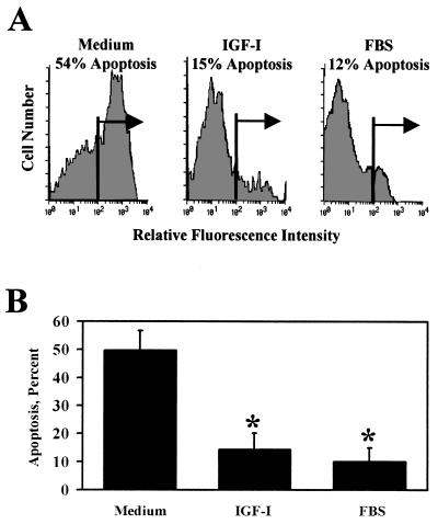
Figure 2.
IGF-I inhibits apoptosis of cerebellar neurons, as determined by intracellular flow cytometry with terminal deoxynucleotide transferase-mediated d-UTP nick-end labeling (TUNEL). (A) Apoptosis of primary cerebellar neurons was measured in granule neurons cultured in medium, IGF-I (100 ng/ml), or 10% FBS with 25 mM KCl. A representative fluorescence histogram shows that treatment with IGF-I reduced the apoptotic cell population from 54% to 15%. Only 12% of neurons cultured with FBS underwent apoptosis. (B) Summary of three independent experiments with flow cytometry using TUNEL. Treatment with IGF-I reduced the apoptotic cell population from 51% ± 8% to 14% ± 4% (P < 0.01, n = 3), and similar results were observed with FBS.

