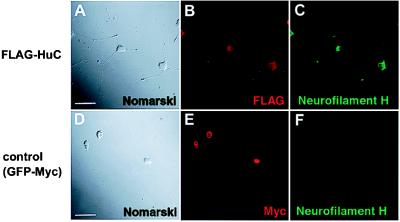
Figure 1.
HuC-induced neuronal phenotype of PC12 cells. (A) Morphology of pCXN2-FLAG-HuC-transfected PC12 cells (Nomarski differential interference contrast optics). Neuron-like morphology, similar to NGF-induced differentiation, appeared 9 d after transfection. (D–F) Negative control (pCXN2-GFP-Myc transfected PC12 cells) showed no morphological changes 9 d after transfection. Double staining for overexpressed FLAG-HuC (or GFP-Myc) (B and E) and neurofilament H (C and F). Antibodies for tagged sequence were used for the detection of transfected fusion protein. Overexpressed HuC protein localized mainly to the cytoplasm, and these cells showed increased expression of Neurofilament H (C), which is also known to increase in the NGF-induced differentiation of PC12 cells (53). (Bar = 15 μm.)

