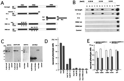
Figure 2.
Mutation analyses of Hu (HuB and HuC) proteins. (A) Mutants of Hu proteins used for functional analysis. HuB and C deletion mutants R1–2 and R3, and HuC RRM amino acid replaced mutants (RRM1mt, RRM2mt) were constructed as described in Material and Methods. (B) RNA-binding assay of HuC mutants. Full-length and mutant HuC fusion proteins and a control fusion protein were incubated with poly(rG)-, poly(rA)-, poly(rU)-, and poly(rC)-Sepharose RHP beads in either 0.25 M, 0.5 M, or 0.8 M NaCl. After being washed, protein bound to the RHP was analyzed by Western blot analysis with an anti-T7 antibody (Novagen). (C) Expression of exogenous and endogenous Hu proteins in PC12 cells 2 d after transfection. To detect these proteins, immunoblot analyses were performed. The expression level of exogenous FLAG-tagged full- length transgene products (indicated by double arrowheads) were quantitated in comparison to the endogenous Hu proteins of control cells (pCXN2 transfected PC12 cells) (indicated by arrowhead). The expression level of Myc-tagged HuB/C-R3 transgene products was indirectly quantitated by comparing to those of Myc-tagged full length transgene product (indicated by small arrow) and endogenous Hu proteins in the control cells. (D) Neuronal differentiation-inducing activities of PC12 cells by Hu mutants. The differentiation-inducing activities of mutants were evaluated by observing the morphology of transfected PC12 cells. G418 was added to the medium to enrich Hu-transfected cells. The cells that had dendrites longer than the diameter of their cell bodies were termed “neuronal phenotype” cells. This graph shows the proportion of differentiated cells among the transgene-containing cells (detected by using antibodies against the FLAG or Myc tags) 9 d after transfection. FLAG-GFP fusion protein was used as a negative control. One hundred transgene-expressing cells were examined three times (total 300 cells) per construct. (E) Hu-R3 mutants act as dominant-negative forms of Hu. Full-length Hu and Hu-R3 mutants were cotransfected into PC 12 cells and analyzed by using the same criteria as in D (single positive and double positive, each 100 cells, three times). Differentiation of cells transfected with both full-length Hu and the Hu-R3 mutant (double-positive cells) was reduced compared with cells transfected with full-length Hu alone (single-positive cells). Transfection of HuC with HuB-R3 or HuB with HuC-R3 showed the same results. Negative controls (pCXN2-FLAG-HuC with pCXN2-GFP-Myc) did not show any inhibition of neuronal differentiation.

